
Abstract
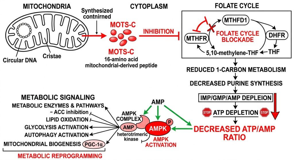
Biomedical consensus traditionally viewed the mitochondrial genome strictly as the blueprint for the essential protein subunits of the oxidative phosphorylation system. The discovery of MOTS-c (Mitochondrial Open Reading Frame of the 12S rRNA type-c) shattered this paradigm. Encoded entirely within a short open reading frame of the mitochondrial 12S rRNA gene, this 16-amino acid peptide acts as an endocrine and intracrine signaling molecule. In advanced in vitro cellular models—primarily utilizing skeletal muscle myocytes and hepatic cellular lines—synthetic MOTS-c demonstrates profound regulatory control over cellular metabolism. This comprehensive review dissects the unique biophysical mechanism by which MOTS-c deliberately induces metabolic stress through targeted inhibition of the one-carbon folate cycle, leading directly to the potent activation of the master metabolic sensor, AMP-activated protein kinase (AMPK).

1. Introduction: Unveiling the Mitochondrial Proteome
Mitochondria are universally recognized as cellular power plants, responsible for the vast majority of ATP synthesis via the electron transport chain. However, their role as complex signaling organelles is coming into sharper focus.
The mitochondrial genome (mtDNA) is small, circular, and heavily optimized. Historically, “junk” or non-coding regions were ignored. Advanced ribosomal profiling eventually revealed that short transcripts within the 12S rRNA gene were actively translated into functional micropeptides. MOTS-c was identified as a primary metabolic regulator originating from this newly mapped mitochondrial micro-proteome.
In in vitro applications, synthetic MOTS-c is highly sought after by researchers seeking to modulate the “exercise mimetic” pathways independently of physical cellular stressors (like hypoxia or forced contractile stimulation).
2. The Unique Intracrine Mechanism: Folate Cycle Inhibition
Conventional activators of cellular metabolism generally operate by directly binding kinase receptors on the cell surface. MOTS-c operates through a fundamentally distinct, intracellular inhibitory mechanism.
Upon in vitro administration to isolated human skeletal muscle cells (myoblasts/myotubes), MOTS-c rapidly translocates. While a fraction exerts paracrine/endocrine effects externally, a significant portion operates intracrinally. The primary cytoplasmic target of MOTS-c is the one-carbon folate cycle.
The folate cycle is a critical metabolic hub responsible for the de novo synthesis of purines (adenine and guanine), which form the structural backbone of ATP. In vitro isotopic tracing studies demonstrate that MOTS-c specifically binds to and inhibits enzymes within this cycle.
By strategically blocking the one-carbon metabolic flux, MOTS-c starves the cell of the precursors necessary to synthesize new purines. This causes an abrupt, highly controlled collapse in de novo ATP synthesis.
3. AICAR Accumulation and AMPK Phosphorylation
The induced failure of purine synthesis creates a cascade effect that perfectly mimics acute cellular energy stress (analogous to profound physical exertion or caloric restriction).
3.1 The Rise of the AMP:ATP Ratio
As the cell continues to consume existing ATP for basic survival functions, but fails to synthesize new ATP due to the MOTS-c mediated folate cycle blockade, the intracellular ratio of Adenosine Monophosphate (AMP) to Adenosine Triphosphate (ATP) spikes dramatically.
Furthermore, the inhibition of the folate pathway causes the upstream accumulation of an intermediate metabolite: 5-aminoimidazole-4-carboxamide ribonucleotide (AICAR). AICAR is an AMP analog and a classically recognized pharmacological activator of AMPK.
3.2 Activation of the Master Switch (AMPK)
AMP-activated protein kinase (AMPK) exists as a heterotrimeric complex ($ alpha, beta, gamma $ subunits) functioning as the cell’s ultimate fuel gauge. When AMP/ATP ratios climb precipitously, AMP binds to the $gamma$-subunit of AMPK. This binding causes a conformational change that exposes a specific threonine residue (Thr172) on the catalytic $alpha$-subunit.
In in vitro western blot analysis of MOTS-c treated cells, researchers observe rapid and intense phosphorylation at Thr172 by upstream kinases (like LKB1). This phosphorylation fully activates the AMPK enzyme.
4. Downstream Metabolic Consequences in Vitro
Once activated by MOTS-c, AMPK initiates a sweeping, cell-wide survival protocol designed to restore energy homeostasis by aggressively ramping up catabolic (energy-producing) pathways and shutting down anabolic (energy-consuming) processes.
- GLUT4 Translocation and Glucose Uptake: In isolated skeletal muscle models, activated AMPK bypasses the insulin-dependent pathway entirely, forcibly recruiting GLUT4 transport vesicles from intracellular storage pools. These vesicles fuse with the plasma membrane, drastically increasing the influx of extracellular glucose into the cell. This represents a primary mechanism of insulin sensitization.
- Hepatic Gluconeogenesis Suppression: When MOTS-c is applied to in vitro hepatic (liver) cell models, AMPK phosphorylates and inhibits critical transcription factors governing gluconeogenesis (the production of new glucose). This effectively shutters the liver cell’s ability to release glucose, a crucial pathway of interest for countering metabolic syndrome profiles.
- Lipid Oxidation via ACC Inhibition: AMPK directly phosphorylates and turns off Acetyl-CoA Carboxylase (ACC). The inhibition of ACC stops the synthesis of malonyl-CoA. Because malonyl-CoA normally inhibits CPT-1 (the gatekeeper enzyme that allows fatty acids to enter the mitochondria for burning), dropping malonyl-CoA levels essentially opens the floodgates. In vitro, MOTS-c treated cells shift rapidly from relying on glucose to aggressively oxidizing intracellular lipid stores via mitochondrial $beta$-oxidation.
5. Nuclear Translocation and Gene Expression
Beyond cytosolic kinase activity, synthetic MOTS-c exhibits a remarkable secondary function under conditions of intense metabolic stress. In vitro models utilizing glucose deprivation show that MOTS-c physically translocates into the cellular nucleus.
Once inside the nucleus, MOTS-c acts as a transcriptional co-regulator. It specifically targets DNA sequences known as Antioxidant Response Elements (AREs). By binding near these sites, MOTS-c orchestrates the upregulation of a vast suite of protective, antioxidant, and survival genes, essentially rewiring the cell’s genomic response to physiological stress.
6. Conclusion
The synthetic peptide MOTS-c provides a fascinating, highly precise tool for in vitro researchers. By acting as a deliberate saboteur of the folate cycle, it engineers a localized purine famine and AICAR accumulation. This precisely mimics profound energetic stress, rapidly flipping the AMPK switch to “on.” The resulting cascade—massive glucose uptake independent of insulin, aggressive lipid oxidation, and adaptive nuclear gene transcription—cements MOTS-c as a profoundly powerful modulator of cellular metabolic destiny and an indispensable asset in modern endocrinological modeling.
Scientific References & Further Reading:
- Lee, C., et al. (2015). The mitochondrial-derived peptide MOTS-c promotes metabolic homeostasis and reduces obesity and insulin resistance. Cell Metabolism, 21(3), 443–454.
- Kim, S. J., et al. (2018). The mitochondrial-derived peptide MOTS-c promotes metabolic homeostasis and reduces obesity and insulin resistance. Journal of Molecular Endocrinology, 60(3), R125-R136.
- Zhai, D., et al. (2017). MOTS-c peptide increases survival and decreases bacterial load in mice infected with MRSA. Molecular Immunology, 92, 151-160.
- Reynolds, J.C., et al. (2021). MOTS-c: A mitochondrial signal regulating metabolism and aging. Frontiers in Endocrinology, 12, 678778.
(Disclaimer: The content detailed above is intended strictly for in vitro laboratory research and academic reference. Synthetic peptides discussed herein are not approved, designed, or strictly evaluated for human consumption, diagnostics, or therapeutic interventions.)