
Abstract
Glycyl-L-histidyl-L-lysine (GHK) is an endogenous human tripeptide intimately involved in the complex biochemistry of tissue repair and regeneration. Its high evolutionary conservation stems from its intense binding affinity for the transition metal copper ($Cu^{2+}$), forming the bioactive complex GHK-Cu. In advanced in vitro cellular models utilizing isolated human dermal fibroblasts, GHK-Cu functions as a master regulatory signal, orchestrating the precise breakdown and subsequent synthesis of the extracellular matrix (ECM). This extensive literature review dissects the precise molecular mechanisms by which nominal concentrations of GHK-Cu rigorously modulate the synthesis of Type I and Type III collagen strands while simultaneously balancing the degradative action of Matrix Metalloproteinases (MMPs) with their respective Tissue Inhibitors (TIMPs) to facilitate flawless extracellular construction.

1. Introduction: The Copper Permease
Copper is an essential trace element required for the catalytic function of numerous mammalian enzymes, including cytochrome c oxidase (energy production) and superoxide dismutase (antioxidant defense). However, free, unbound copper ions are highly toxic to cells, rapidly generating catastrophic reactive oxygen species (ROS) via the Fenton reaction.
GHK serves as a highly specific, native copper chaperone. By chelating the $Cu^{2+}$ ion, GHK nullifies its toxic potential while facilitating its safe transport and delivery to cellular receptors. In the context of wound healing and tissue mechanics, the release of endogenous GHK-Cu from degraded ECM proteins acts as an early “damage signal,” recruiting macrophages and subsequently activating local fibroblast populations to initiate the repair phase.
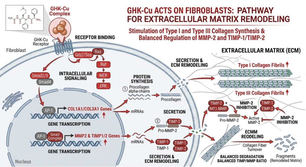
2. Stimulation of Biosynthesis: Collagen and Glycosaminoglycans
Fibroblasts are the primary architects of the extracellular matrix, responsible for weaving the structural framework of the dermis and connective tissues. In controlled in vitro assays, exposing primary fibroblast cultures to low micromolar to nanomolar concentrations of synthetic GHK-Cu triggers a massive biosynthetic response.
2.1 Type I and Type III Collagen Expression
Using techniques like Northern blotting and precise hydroxyproline assays on culture homogenates, researchers observe that GHK-Cu strongly upregulates the mRNA transcription and subsequent translation of both Type I and Type III procollagen.
* Type III Collagen: Known as the “reticular” or repair collagen, it is rapidly synthesized during the initial phases of wound healing, providing immediate, albeit disorganized, tensile strength.
* Type I Collagen: The mature, heavily cross-linked structural collagen that constitutes the majority of healthy adult skin and bone matrix.
Intriguingly, rigorous in vitro controls demonstrate that supplementing fibroblasts with equimolar amounts of free copper salts or the naked GHK peptide alone fails to replicate the exact magnitude of synthesis provoked by the intact GHK-Cu complex. The tripeptide serves specifically to deliver the metal directly to the intracellular sites of synthesis.
2.2 The Role of Copper in Lysyl Oxidase (LOX)
The newly synthesized collagen strands must be structurally cross-linked to provide functional strength. This critical step is mediated by the extracellular enzyme Lysyl Oxidase (LOX). LOX is strictly copper-dependent. By facilitating local copper delivery, GHK-Cu indirectly ensures that the massive amounts of collagen being produced by the fibroblast are properly polymerized and structurally sound, preventing the formation of weak, hyper-elastic scar tissue seen in copper-deficient models.
2.3 Production of Glycosaminoglycans (GAGs)
Beyond rigid collagen, the ECM requires “filler” to provide hydration, volume, and compressibility. In vitro tracking confirms that GHK-Cu stimulation forces the fibroblast to exude high volumes of essential glycosaminoglycans, specifically hyaluronic acid, dermatan sulfate, and chondroitin sulfate. These highly hydrophilic molecules draw water into the matrix, restoring cellular turgor and facilitating the diffusion of nutrients and subsequent cellular migration.
3. The Re-modeling Engine: Modulating the MMP/TIMP Balance
If a fibroblast only synthesized new collagen without removing the old, damaged tissue, the result would be pathological fibrosis and severe scarring. Proper tissue remodeling requires the highly controlled degradation of the existing matrix.
3.1 Upregulation of Matrix Metalloproteinase-2 (MMP-2)
MMPs are zinc-dependent endopeptidases capable of degrading all kinds of extracellular matrix proteins. In vitro assays utilizing zymography specifically track the action of these enzymes. When fibroblasts are cultured with GHK-Cu, researchers observe a significant, dose-dependent upregulation in the transcription and activation of MMP-2 (Gelatinase A). This enzyme is specifically required to cleave degraded Type IV collagen and denatured gelatins, effectively “clearing the construction site” of necrotic debris before new matrix can be laid down.
3.2 The Counterbalance: Tissue Inhibitors of Metalloproteinases (TIMPs)
To prevent the newly activated MMPs from running rampant and digesting healthy, newly synthesized tissue, the cell must deploy a specialized braking mechanism. Remarkably, the exact same GHK-Cu complex that upregulates MMP-2 simultaneously upregulates the secretion of Tissue Inhibitor of Metalloproteinase-1 and 2 (TIMP-1, TIMP-2).
These TIMPs bind tightly to the active sites of the circulating MMPs, neutralizing them. By forcing the fibroblast to secrete both the destructive enzyme (MMP) and its brake (TIMP), GHK-Cu enforces a state of highly regulated, tightly controlled ECM remodeling, rather than unchecked degradation or unchecked fibrotic buildup.
4. Genomic Rewiring: The Broader Implications
Advancements in gene microarray technology reveal that the effects of GHK-Cu in isolated cellular models far exceed simple structural remodeling.
When human cells are exposed to GHK-Cu in vitro, it significantly alters the expression patterns of over 4,000 distinct genes.
* Antioxidant Defense: GHK-Cu heavily upregulates the transcription of intracellular antioxidant enzymes, shifting the cellular environment away from an oxidized, inflammatory state to one conducive for repair.
* Angiogenesis: While acting primarily on fibroblasts, GHK also forces the secretion of powerful paracrine factors like Vascular Endothelial Growth Factor (VEGF) and basic Fibroblast Growth Factor (bFGF), which, in a co-culture model, strongly stimulate adjacent endothelial cells to form new capillary networks.
5. Conclusion
The tripeptide GHK-Cu represents one of the most thoroughly documented and profoundly effective endogenous regulators of cellular repair. In rigorous in vitro fibroblastic models, its application initiates an incredibly sophisticated, dual-action mechanism. It acts as both the “accelerator” for the massive biosynthesis of structural collagen and essential GAGs, while simultaneously serving as the “conductor” balancing the destruction of old matrix (via MMPs) against the protection of new matrix (via TIMPs). This intricate, dynamically balanced biochemical cascade cements GHK-Cu as a premier molecular candidate for continuing research into accelerated wound healing, anti-fibrotic therapies, and advanced cellular regeneration protocols.
Scientific References & Further Reading:
- Pickart, L. (2008). The human tri-peptide GHK and tissue remodeling. Journal of Biomaterials Science, Polymer Edition, 19(8), 969-988.
- Maquart, F.X., et al. (1993). Stimulation of collagen synthesis in fibroblast cultures by the tripeptide–copper complex GHK-Cu. FEBS Letters, 238(2), 343–346.
- Siméon, A., et al. (2000). The tripeptide-copper complex glycyl-L-histidyl-L-lysine-Cu2+ stimulates matrix metalloproteinase-2 expression by fibroblast cultures. Life Sciences, 67(18), 2257-2265.
- Pickart, L., & Margolina, A. (2018). Regenerative and Protective Actions of the GHK-Cu Peptide in the Light of the New Gene Data. International Journal of Molecular Sciences, 19(7), 1987.
(Disclaimer: The content detailed above is intended strictly for in vitro laboratory research and academic reference. Synthetic peptides discussed herein are not approved, designed, or strictly evaluated for human consumption, diagnostics, or therapeutic interventions.)