
—
Receptor Pharmacology: In-Vitro Binding and Transduction Mechanisms of Melanocortin Receptor Agonists
Introduction to the Melanocortin System
The central and peripheral melanocortin systems constitute a highly complex and pervasive neuroendocrine signaling network. This system is critically involved in modulating a vast array of physiological processes, including pigmentation (melanogenesis), energy homeostasis, feeding behavior, sexual function, and robust modulation of inflammatory and immune responses. Endogenous melanocortin ligands, such as $alpha$-melanocyte-stimulating hormone ($alpha$-MSH), adrenocorticotropic hormone (ACTH), and various other cleavage products of the pro-opiomelanocortin (POMC) precursor protein, exert their pleiotropic effects by binding to and activating a specific family of receptors. Research focused on synthetic melanocortin agonists (e.g., Melanotan I, Melanotan II, PT-141) necessitates a rigorous understanding of molecular binding affinities, receptor subtype selectivity, and downstream cellular signaling cascades. This analytical review examines the in-vitro pharmacology and receptor-binding characteristics of melanocortin agonists.
The Melanocortin Receptor (MCR) Family Architecture
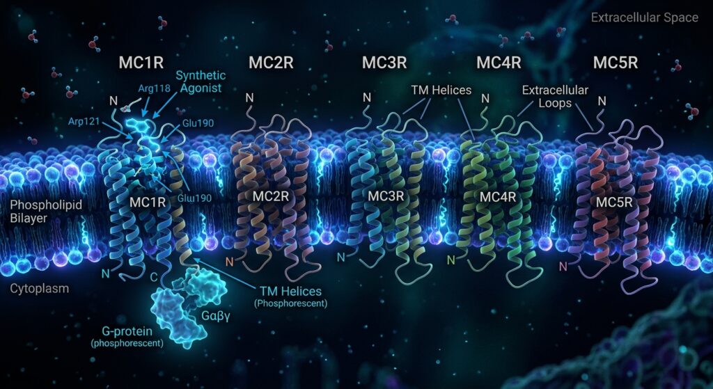
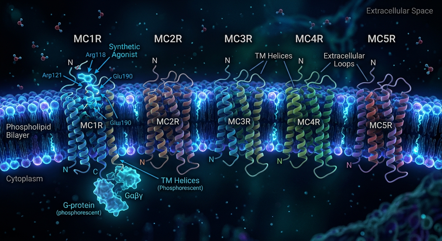
The biological targets of melanocortin peptides are the melanocortin receptors (MCRs), a distinct subfamily of Class A (rhodopsin-like) G-protein-coupled receptors (GPCRs). To date, five distinct receptor subtypes (MC1R through MC5R) have been cloned and structurally characterized. They are distinguished by their specific tissue distribution patterns and their differential affinities for both endogenous ligands and highly specific synthetic analogs.
Structurally, all five MCR subtypes share the classic GPCR topology: seven hydrophobic transmembrane (TM) $alpha$-helices connected by three extracellular and three intracellular loops, with an extracellular N-terminus and an intracellular C-terminus. The binding pocket for melanocortin ligands is deeply localized within the transmembrane bundle, formed primarily by specific highly conserved residues within TM2, TM3, TM6, and TM7.
- MC1R: Predominantly expressed on dermal melanocytes and various leukocyte populations. It is the primary regulator of eumelanin synthesis (pigmentation) and exhibits significant anti-inflammatory signaling capacities.
- MC2R: Uniquely expressed in the adrenal cortex; it strictly binds ACTH (requiring the melanocortin-2 receptor accessory protein, MRAP, for functional surface expression) to rapidly stimulate glucocorticoid steroidogenesis. It generally does not bind MSH analogs.
- MC3R & MC4R: These are the primary “central” receptors, largely localized within the central nervous system (specifically the hypothalamus). They serve as paramount regulators of energy balance, feeding behaviors (anorexigenic signals), and metabolic autonomic functions.
- MC5R: Widely distributed in peripheral exocrine tissues (e.g., sebaceous glands, lacrimal glands), primarily modulating exocrine secretion, though its full functional repertoire is continually expanding.
In-Vitro Pharmacodynamics: Binding Affinity and Selectivity
Investigating synthetic melanocortin agonists in-vitro heavily utilizes competitive radioligand binding assays. In these protocols, precisely cultivated cell lines (such as HEK-293 or CHO cells) are stably transfected to overexpress one specific human MCR subtype. Membrane preparations from these cells are incubated with a fixed concentration of a highly potent radioactive ligand (e.g., $[^{125}I]$-NDP-$alpha$-MSH) and increasing, graded concentrations of the novel synthetic peptide under structural investigation.
The synthetic agonist competes with the radioligand for the specific binding pocket. By quantifying the displacement of radioactivity, researchers calculate the Inhibition Constant ($K_i$), which represents the absolute affinity of the synthetic peptide for that specific receptor subtype. A lower $K_i$ value denotes higher binding affinity.
Endogenous $alpha$-MSH is a non-selective agonist, binding with approximately equal, high affinity to MC1R, MC3R, MC4R, and MC5R. Synthetic research peptides are often structurally engineered to alter this selectivity profile.
For example, the synthetic analogue Melanotan II (MT-II, a cyclic heptapeptide lactam: Ac-Nle-cyclo[Asp-His-D-Phe-Arg-Trp-Lys]-$NH_2$) incorporates structural constraints (cyclization) and specific amino acid substitutions (Norleucine for Methionine, D-Phenylalanine for L-Phenylalanine) derived from the endogenous $alpha$-MSH sequence. In-vitro binding data demonstrates that MT-II is a highly potent, exceptionally stable, but fundamentally non-selective pan-agonist. It exhibits powerful low-nanomolar binding affinities ($K_i$) across MC1R, MC3R, MC4R, and MC5R. This lack of profound selectivity explains its broad-spectrum biological manifestations in research models, including simultaneous potent melanogenesis (MC1R) and central metabolic/arousal effects (MC3R/MC4R).
Conversely, other synthetic efforts focus on strict receptor isolation. Agonists heavily restricted to activating only MC1R are sought for specific dermatological or anti-inflammatory cellular assays without triggering the anorexigenic side effects associated with central MC4R activation.
Signal Transduction: The cAMP Intracellular Cascade
Upon successful binding to the extracellular domain/transmembrane pocket of a melanocortin receptor, the agonist stabilizes an active conformational state of the GPCR. This structural shift allows the intracellular loops of the receptor to positively couple with and activate specific heterotrimeric G-proteins.
All five established melanocortin receptors primarily couple to the stimulatory G-protein, $Galpha_s$. In-vitro evaluation of functional receptor activation (agonism versus antagonism) relies heavily on measuring the downstream consequences of this specific G-protein coupling.
Upon agonist activation, the $Galpha_s$ subunit rapidly dissociates from the $Gbetagamma$ dimer and profoundly stimulates the membrane-bound effector enzyme, adenylyl cyclase (AC). Activated adenylyl cyclase catalyzes the rapid, massive conversion of intracellular adenosine triphosphate (ATP) into the critical ubiquitous second messenger molecule, cyclic adenosine monophosphate (cAMP).
In-vitro functional assays directly quantify this intracellular cAMP accumulation (often using time-resolved fluorescence resonance energy transfer, TR-FRET, or enzyme-linked immunosorbent assays, ELISA) in response to varying agonist concentrations. This data generates dose-response curves, allowing for the precise calculation of the half-maximal effective concentration ($EC_{50}$). The $EC_{50}$ defines the potency of the agonist in triggering the primary cellular response. A highly potent agonist will trigger maximal cAMP generation at extremely low (picomolar to low nanomolar) concentrations.
Downstream Kinase Activation and Cellular Functional Assays
The massive surge in intracellular cAMP triggered by MCR agonism is not the endpoint; it acts as a rapid initiation signal. The primary direct target of elevated cAMP is Protein Kinase A (PKA). cAMP binds specifically to the regulatory subunits of inactive PKA tetramers, forcing a conformational change that releases and fully activates the catalytic subunits.
Activated PKA subsequently rapidly phosphorylates a massive cascade of specific downstream protein targets, dictating the ultimate cellular response based on the specific cell type.
* In Melanocyte Models (MC1R): In-vitro assays on cultured B16 melanoma cells demonstrate that sustained PKA activation upregulates the transcription factor MITF (Microphthalmia-associated transcription factor), fundamentally driving the expression of tyrosinase, the rate-limiting enzyme required for the synthesis of complex melanin pigments. Functional assays frequently quantify this resulting tyrosinase activity or total melanin output.
* In Macrophage Models (MC1R/MC3R): In models of induced cellular inflammation (e.g., LPS-stimulated RAW 264.7 macrophages), melanocortin agonism triggers complex intracellular inhibitory pathways. High cAMP levels influence NF-$kappa$B (Nuclear Factor kappa-light-chain-enhancer of activated B cells) signaling, preventing its nuclear translocation. In-vitro, this is quantified by a rapid, profound dose-dependent reduction in the secretion of pro-inflammatory cytokines such as TNF-$alpha$, IL-6, and Nitric Oxide (NO) into the culture media.
Conclusion
The characterization of synthetic melanocortin agonists for research purposes requires stringent in-vitro analytical evaluation. Understanding the nuanced interplay between specific molecular structural modifications, subsequent precise binding affinities ($K_i$) across the differing MCR subtypes, and the resulting quantitative potentiation of the adenylate cyclase/cAMP/$Galpha_s$ signal transduction cascade ($EC_{50}$) is crucial. These robust in-vitro metrics define the unique pharmacological profile of experimental melanocortin ligands prior to complex organismal evaluation.