

Abstract
The structural and biochemical characterization of synthetic polypeptides is paramount in advanced in vitro laboratory environments. While traditional sequencing modalities provided basic structural data, the advent and refinement of Mass Spectrometry (MS) have revolutionized the capacity to identify, characterize, and sequence peptides with unparalleled resolution. By converting molecules into charged ions and meticulously analyzing their mass-to-charge ($m/z$) ratios, modern MS methodologies allow for profound insights into molecular weights, exact amino acid sequencing, and intricate post-translational modifications (PTMs). This technical review evaluates the primary MS techniques—including MALDI-TOF, ESI, and LC-MS/MS—and dissects their critical roles in ensuring the fidelity and stability of peptides during controlled in vitro experimentation.
1. Introduction to High-Resolution Mass Spectrometry (HRMS) Dynamics
Proteins and their smaller peptide derivatives dictate nearly all cellular function through highly specific structural interactions. In the context of in vitro research, securing absolute certainty over a peptide’s primary sequence and its structural integrity is the critical first step before any cell-culture or binding-affinity assay can commence.
Mass spectrometry functions by ionizing chemical species and sorting the ions based on their $m/z$ ratio. In peptide science, this process begins with the vaporization and ionization of the sample. For large biomolecules like peptides, soft ionization techniques are strictly required to prevent the complete fragmentation and destruction of the molecule prior to analysis.
The resulting spectra yield highly accurate molecular weights. When coupled with tandem mass spectrometry (MS/MS), these parent ions are further fragmented in a controlled manner, allowing researchers to piece together the exact sequence of amino acids, much like reconstructing a shattered puzzle from its fragments. This level of precision is indispensable for verifying synthetic peptide batches, distinguishing enantiomers, and mapping complex peptide-receptor interactions in isolated cellular models.
2. Fundamental Ionization Methodologies in Analytical Chemistry
The transition of a peptide from a solid or liquid state into gas-phase ions without significant thermal degradation represents a complex biophysical challenge. Two primary “soft ionization” techniques dominate the field of advanced peptide research.
2.1 Matrix-Assisted Laser Desorption/Ionization (MALDI)
MALDI has long stood as a robust methodology for rapid molecular mass determination. In this technique, the peptide target is co-crystallized with a large excess of a matrix compound (often weak organic acids like $alpha$-cyano-4-hydroxycinnamic acid) on a metal target plate.
A pulsed laser (commonly a nitrogen laser at 337 nm) irradiates the spot. The matrix preferentially absorbs the laser energy, facilitating rapid sublimation. This phase transition carries the intact peptide molecules into the gas phase. During this expansion, proton transfer occurs from the matrix to the peptide, generating predominantly singly charged pseudo-molecular ions $[M+H]^+$.
MALDI is frequently coupled with Time-of-Flight (TOF) analyzers (MALDI-TOF). Given its high tolerance for salts and buffers often used in biological preparations, MALDI-TOF is rigorously utilized for high-throughput batch verification and purity assessment of synthesized peptides in vitro.
2.2 Electrospray Ionization (ESI)
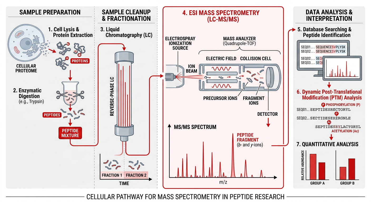
Unlike MALDI, Electrospray Ionization constructs ions directly from solution, making it intrinsically suited for coupling with upstream liquid separation techniques.
In ESI, a peptide solution is pumped through a fused-silica capillary. A high voltage (typically 2-6 kV) is applied to the capillary tip. As the liquid exits, it forms a Taylor cone under the influence of the strong electric field, emitting a fine aerosol of highly charged droplets. As solvent rapidly evaporates—often assisted by a heated drying gas—the charge density on the droplet surface increases until it reaches the Rayleigh limit. At this critical juncture, Coulombic repulsion overcomes surface tension, resulting in droplet fission. This process repeats until bare, multiply charged gas-phase peptide ions $[M+nH]^{n+}$ are released into the vacuum of the mass spectrometer.
Because ESI generates multiply charged ions, it brings large, massive polypeptides into the $m/z$ range of standard analyzers (like quadrupoles or ion traps), drastically expanding the analyzable mass range and facilitating superior fragmentation in MS/MS experiments.
3. Advanced Analyzers and Tandem Mass Spectrometry (MS/MS)
Securing the precise mass of a peptide is critical for structural confirmation, but determining the exact amino acid sequence—the primary structure—requires tandem mass spectrometry, or MS/MS.
3.1 Liquid Chromatography–Tandem Mass Spectrometry (LC-MS/MS)
In complex in vitro environments, identifying a single peptide profile amongst thousands of background cellular proteins necessitates dynamic separation. High-Performance Liquid Chromatography (HPLC) or Ultra-High-Performance Liquid Chromatography (UHPLC) coupled downstream to an ESI-MS system represents the gold standard.
Peptides are initially separated in the liquid phase based on hydrophobicity, typically utilizing a reverse-phase column (e.g., C18). As they elute incrementally into the ESI source, the first mass analyzer (MS1) isolates a specific intact peptide precursor ion of interest.
3.2 Collision-Induced Dissociation (CID) and Peptide Sequencing
Once isolated, the precursor ion is subjected to targeted fragmentation, most commonly via Collision-Induced Dissociation (CID) or Higher-energy Collisional Dissociation (HCD). The isolated peptide ion is accelerated into a collision cell containing an inert neutral gas (like argon or nitrogen). Repeated collisions convert kinetic energy into internal vibrational energy, eventually causing the peptide to fracture.
Due to the fundamental structure of peptides, fragmentation in CID/HCD predominantly occurs along the weakest bond—the amide bond along the peptide backbone. This specific cleavage generates characteristic “b-ions” (charge retained on the N-terminal fragment) and “y-ions” (charge retained on the C-terminal fragment).
The secondary mass analyzer (MS2) then measures these fragments. By calculating the mass differences between sequential b-ions or sequential y-ions, the exact amino acid sequence can be mathematically deciphered. This is mathematically rigorous and allows for definitive verification that synthetic sequences match their intended design prior to deployment in sensitive in vitro cellular assays.
4. Crucial In Vitro Applications of MS in Peptide Science
The deployment of advanced HRMS frameworks facilitates a wide array of in vitro investigations:
- Absolute Structural Validation: Prior to evaluating a peptide’s affinity toward a receptor (e.g., G-Protein Coupled Receptors in cell lysates), exact primary structure validation via LC-MS/MS prevents costly errors resulting from synthetic truncation or omission.
- Identification of Post-Translational Modifications (PTMs): Physiological peptides rarely act in their naked form. Modifications such as phosphorylation, acetylation, or glycosylation dictate receptor binding kinetics. MS/MS can pinpoint the exact chemical group and the specific amino acid residue modified by observing mass shifts in the fragmentation spectrum.
- Receptor Cross-Linking and Interaction Mapping: Advanced in vitro workflows utilize MS to map exact binding domains. Peptides can be covalently cross-linked to their target receptors in isolated cellular extracts. Subsequent enzymatic digestion and MS analysis of the cross-linked fragments allow investigators to identify the precise contact points, elucidating three-dimensional structural interactions.
- In Vitro Degradation and Stability Profiling: Understanding a peptide’s half-life is critical. Peptides are incubated in specific biological matrices (like human serum homogenates or cell lysate extracts), and samples are taken at timed intervals. Quantitative LC-MS/MS precisely monitors the depletion of the parent peptide and identifies degradation metabolites, illuminating the sites of proteolytic cleavage and guiding the design of more structurally stable analogs.
5. Conclusion
Mass spectrometry remains the foundational pillar of modern peptide characterization. The continuous evolution of ionization methodologies and state-of-the-art analyzers such as the Orbitrap has pushed the boundaries of speed, sensitivity, and mass accuracy. In rigorous in vitro research settings, MS methodologies provide the definitive, high-resolution empirical data necessary to confidently map peptide structures, decipher complex metabolic degradation pathways, and investigate highly specific receptor-ligand interactions at the molecular level.
Scientific References & Further Reading:
- Aebersold, R., & Mann, M. (2003). Mass spectrometry-based proteomics. Nature, 422, 198–207.
- Yates, J.R., et al. (2009). Proteomics by mass spectrometry: approaches, advances, and applications. Annual Review of Biochemistry, 78, 243–272.
- Zubarev, R. A., Kelleher, N. L., & McLafferty, F. W. (1998). Electron Capture Dissociation of Multiply Charged Protein Cations. A Nonergodic Process. Journal of the American Chemical Society, 120(13), 3265–3266.
- Aebersold, R., & Mann, M. (2016). Mass-spectrometric exploration of proteome structure and function. Nature, 537, 347–355.
- Olsen, J. V., et al. (2007). Higher-energy C-trap dissociation for peptide modification analysis. Nature Methods, 4, 709–712.
(Disclaimer: The content detailed above is intended strictly for in vitro laboratory research and academic reference. Synthetic peptides discussed herein are not approved, designed, or strictly evaluated for human consumption, diagnostics, or therapeutic interventions.)