

Abstract
Ipamorelin is a synthetic, pentapeptide growth hormone secretagogue (GHS) originally engineered for enhanced receptor specificity compared to first-generation hexapeptides like GHRP-6 and GHRP-2. Designed to mimic the endogenous ligand ghrelin, Ipamorelin binds exclusively to the Growth Hormone Secretagogue Receptor 1a (GHS-R1a). In targeted in vitro models utilizing isolated pituitary somatotrophs, Ipamorelin demonstrates an unprecedented capacity to trigger the exocytosis of Growth Hormone (GH) vesicles while maintaining profound stoichiometric silence regarding the secondary release of adrenocorticotropic hormone (ACTH), cortisol, and prolactin. This detailed literature synthesis and biological review dissects the receptor binding kinetics, G-protein coupled intracellular signaling cascades, and the profound receptor specificity of Ipamorelin in advanced cellular modeling.
1. Introduction: Evolution of Growth Hormone Secretagogues
The anterior pituitary gland tightly regulates the synthesis and pulsatile release of Growth Hormone via somatotroph cells. This pulsatility is naturally governed by the opposing forces of Growth Hormone-Releasing Hormone (GHRH, stimulatory) and Somatostatin (inhibitory). The discovery of a third regulatory pathway—the ghrelin/GHS-R1a axis—revolutionized neuroendocrine biology.
Early synthetic ligands for this receptor, such as GHRP-6, demonstrated potent capability to induce GH secretion in vitro. However, they were universally plagued by “off-target” receptor promiscuity resulting in the undesired synthesis and release of stress hormones, specifically cortisol and ACTH. Ipamorelin (Aib-His-D-2-Nal-D-Phe-Lys-NH2) was synthesized with a specific focus on structural modification—specifically the incorporation of the $alpha$-aminoisobutyric acid (Aib) and D-2-naphthylalanine (D-2-Nal) residues—to severely restrict its binding affinity strictly to the GHS-R1a receptor, rendering it highly selective.
2. Receptor Binding and G-Protein Transduction Kinetics
In vitro binding assays utilizing radiolabeled homologous displacement techniques confirm that Ipamorelin possesses an extraordinarily high binding affinity for the cloned GHS-R1a receptor, possessing a $K_i$ (inhibition constant) typically ranging dynamically in the low nanomolar brackets. The GHS-R1a is a classic seven-transmembrane domain G-Protein Coupled Receptor (GPCR).
2.1 Activation of the $G_{alpha q/11}$ Subunit
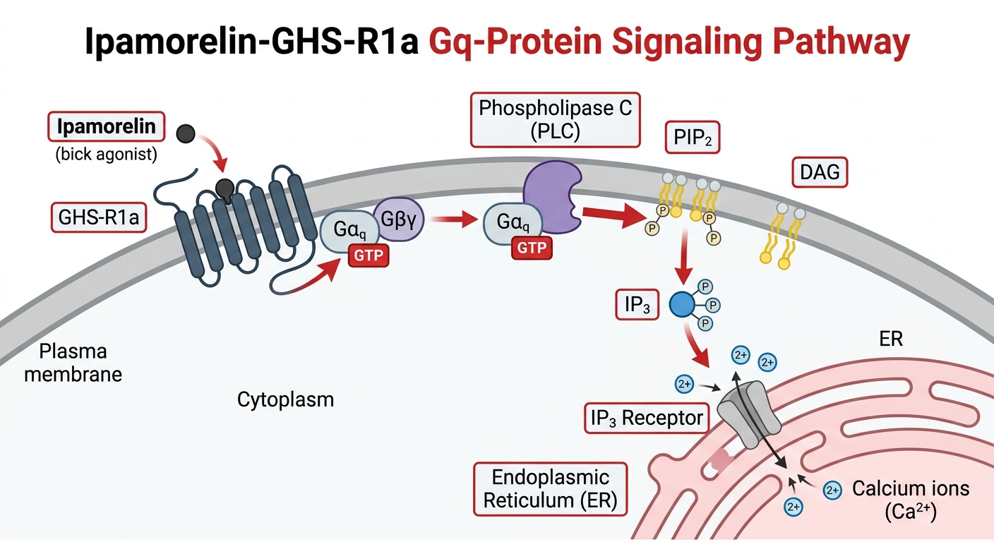
Unlike GHRH, which binds its receptor and elevates intracellular cyclic AMP (cAMP) via the $G_{alpha s}$ pathway, the binding of Ipamorelin to the GHS-R1a induces a localized conformational shift that recruits and exchanges GDP for GTP on the associated heterotrimeric $G_{alpha q/11}$ protein complex.
This specific G-protein coupling is critical. Once activated in vitro, the $G_{alpha q/11}$ subunit dissociates and translocates along the inner leaflet of the plasma membrane until it encounters its primary effector enzyme: Phospholipase C (PLC).
2.2 The Phosphatidylinositol Pathway: PIP2, IP3, and DAG
Activated Phospholipase C executes a highly specific enzymatic cleavage. It hydrolyzes phosphatidylinositol 4,5-bisphosphate (PIP2), a minor but critical phospholipid component of the cellular membrane, splitting it into two distinct, heavily active second messengers:
- Diacylglycerol (DAG): Remains tethered in the hydrophobic plasma membrane environment where it acts as a docking and activation site for Protein Kinase C (PKC).
- Inositol 1,4,5-trisphosphate (IP3): A water-soluble molecule that diffuses rapidly through the cytoplasm.
2.3 Intracellular Calcium Mobilization
The newly generated IP3 migrates to the rough endoplasmic reticulum (ER) within the somatotroph. Here, it binds to specific IP3 receptors ($IP_3R$), which act as ligand-gated calcium channels. Binding triggers a rapid, massive efflux of stored $Ca^{2+}$ ions from the ER lumen into the cytoplasm.
In in vitro fluorescence microscopy utilizing $Ca^{2+}$-sensitive dyes (like Fura-2), the administration of Ipamorelin to isolated somatotrophs induces a sharp, robust, and immediate “spike” in intracellular free calcium. This calcium spike is the terminal execution signal for exocytosis. It induces specialized SNARE proteins to pull GH-containing secretory vesicles to the plasma membrane, fusing them and expelling Growth Hormone into the extracellular space.
3. The Hallmark of Ipamorelin: Exquisite Receptor Specificity
The fundamental differentiator of Ipamorelin in advanced laboratory research lies not in how it releases GH, but in what it fails to release.
In controlled in vitro cellular assays comprising mixed anterior pituitary cellular populations, first-generation secretagogues like GHRP-2 consistently stimulate secondary receptors, causing corticotrophs and lactotrophs to release massive amounts of ACTH and Prolactin alongside GH.
Conversely, rigorous mass-spectrometry and ELISA profiling of culture media post-Ipamorelin administration indicate a near-zero deviation in baseline ACTH and prolactin levels. The structural constraints inherent in the pentapeptide sequence of Ipamorelin prevent it from agonizing the secondary receptor pathways prevalent in earlier GHRPs. This makes Ipamorelin the premier synthetic secretagogue for in vitro studies seeking to isolate and interrogate the ghrelin/GHS-R1a axis without the confounding variable of a systemic stress-hormone cascade.
4. In Vitro Applications and Future Modeling
Given its absolute specificity, Ipamorelin is extensively deployed in several prominent in vitro study models:
- Pulsatility Dynamics: Researchers utilize continuously perfused pituitary cellular matrices to study how pulsatile exposure to Ipamorelin regulates the desensitization or up-regulation of the GHS-R1a receptor over extended timeframes.
- Synergistic Overlap Mapping: Ipamorelin is frequently co-administered with GHRH analogues in vitro. Because GHRH operates via the cAMP pathway and Ipamorelin via the $IP_3/Ca^{2+}$ pathway, researchers can map the intracellular cross-talk and massive synergistic burst in GH release that occurs when both pathways combine.
- Chondrocyte and Osteoblast Research: Emerging evidence indicates that GHS-R1a is also distributed in peripheral tissues, specifically cartilage and bone matrices. In vitro assays currently evaluate how localized Ipamorelin binding might directly influence chondrocyte proliferation and osteoblastic differentiation independent of the pituitary axis.
5. Conclusion
The pentapeptide Ipamorelin stands as a triumph of targeted, structure-based molecular design. By seamlessly initiating the $G_{alpha q/11}$ linked phosphatidylinositol signaling cascade specifically at the GHS-R1a receptor, it generates massive intracellular calcium mobilization required for robust Growth Hormone secretion in vitro. Crucially, its structurally mandated inability to agonize subsequent receptor classes separates it from predecessor oligopeptides, allowing pure, unadulterated research into the mechanisms of endocrine growth modulation without corrupting the resulting dataset with off-target stress hormone artifacts.
Scientific References & Further Reading:
- Raun, K., et al. (1998). Ipamorelin, the first selective growth hormone secretagogue. Biochemical and Biophysical Research Communications, 250(2), 262–267.
- Smith, R.G., et al. (2000). Receptor-specific actions of novel growth hormone secretagogues. Journal of Clinical Endocrinology & Metabolism, 85(8), 6698–6705.
- Johansen, P. B., et al. (1999). Ipamorelin, a selectively acting growth hormone secretagogue: Pharmacokinetics and pharmacodynamics. Growth Hormone & IGF Research, 9(2), 106-113.
- Lu, C. Y., et al. (2006). The Growth Hormone Secretagogue Receptor. Physiological Reviews, 86(3), 1075-1100.
(Disclaimer: The content detailed above is intended strictly for in vitro laboratory research and academic reference. Synthetic peptides discussed herein are not approved, designed, or strictly evaluated for human consumption, diagnostics, or therapeutic interventions.)